Quantitative Assessment of Lipid Accumulation in Liver and Spleen for LNP Delivery Systems in Basel

PROVEN INTELLIGENCE ACCELERATING NEXT-GENERATION THERAPIES

Quantitative Assessment of Lipid Accumulation in Liver and Spleen for LNP Delivery Systems in Basel

Quantitative Assessment of Lipid Accumulation in Liver and Spleen for LNP Delivery Systems

CELL & GENE | RNA | BIOLOGICS
What specific staining techniques are used for lipid quantification in LNP studies?

Our core platform utilizes Oil Red O (ORO) and Sudan Black B for the visualization and quantification of neutral lipids within cryosections. We supplement these with H&E for morphological context and can deploy custom immunohistochemistry (IHC) protocols to co-localize LNPs with specific cell markers, such as Kupffer cells or splenic macrophages.

How does your digital pathology platform differentiate between intracellular and extracellular lipid accumulation?

We employ advanced image analysis algorithms that use cellular segmentation based on nuclear counterstains (e.g., DAPI or hematoxylin). The software defines cytoplasmic boundaries for each cell, allowing the quantification algorithm to measure lipid droplet area and intensity specifically within those boundaries, thereby distinguishing intracellular from extracellular deposits.

What is the typical sample throughput for a non-target tissue biodistribution study focused on liver and spleen?

For a standard study design, our automated systems can process, stain, and scan up to 200 slides per day. The subsequent quantitative analysis is typically completed within 3-5 business days, providing a comprehensive data package. This capacity supports the high-throughput screening demands of LNP formulation optimization.

Can this quantitative assessment be integrated with other endpoints?

Yes. The histology data is designed for integration. We frequently correlate quantitative lipid accumulation data with parallel analyses, such as qPCR for mRNA payload expression or multiplex immunoassays for cytokine profiling from the same tissue blocks, providing a multi-parametric view of LNP efficacy and safety.

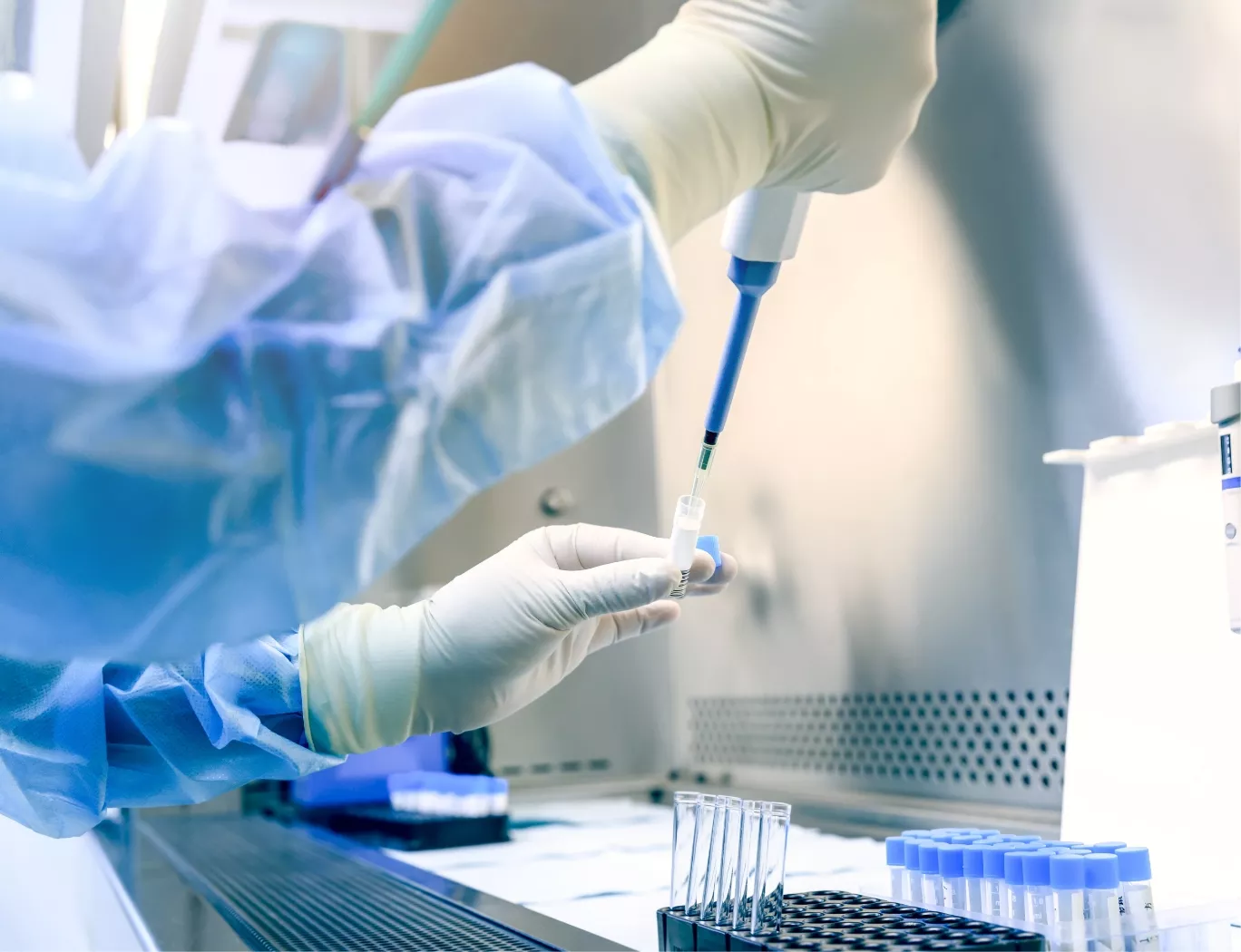
A scientist in protective gear pipetting a sample into a vial within a sterile laboratory hood.

Executive Summary

Optimizing lipid nanoparticle (LNP) delivery requires precise quantification of organ-specific accumulation. Helper lipid composition directly dictates whether an LNP system targets the liver or the spleen, a determining factor for both efficacy and safety profiles. Our GxP-compliant digital pathology platform provides sponsors with high-resolution, quantitative data on lipid biodistribution in key tissues. We replace subjective scoring with objective, algorithm-driven analysis of lipid accumulation, enabling direct comparison of LNP formulations and accelerating the selection of lead candidates for IND-enabling programs.

The Challenge of LNP Biodistribution

The biodistribution of an LNP is a dynamic outcome, determined by its structural components. The selection of helper lipids, for instance, profoundly influences the protein corona that forms around the nanoparticle in vivo. This protein adsorption, particularly of apolipoproteins like ApoE, governs subsequent organ uptake.

Recent investigations highlight this relationship with precision. As demonstrated in high-throughput in vivo screening studies, LNP formulations containing DOPE as a helper lipid show preferential accumulation in the liver, whereas those formulated with DSPC exhibit a distinct tropism for the spleen (PMID: 33404020). This has direct consequences for therapeutic development:

  • Liver-Targeted LNPs: Ideal for therapies addressing hepatic diseases or for applications requiring high levels of protein expression via hepatocyte transfection.

  • Spleen-Targeted LNPs: Advantageous for immunomodulatory applications, vaccine development, or therapies targeting splenic immune cell populations.

Understanding and controlling this differential targeting is fundamental to developing a viable therapeutic. It requires a robust, reproducible method for quantifying lipid accumulation at the cellular level.

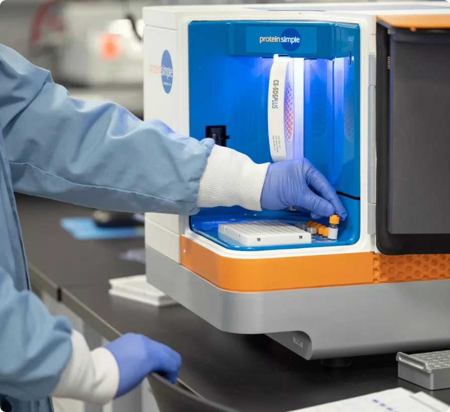
A scientist in a lab coat and gloves loads samples into a ProteinSimple instrument for analysis.

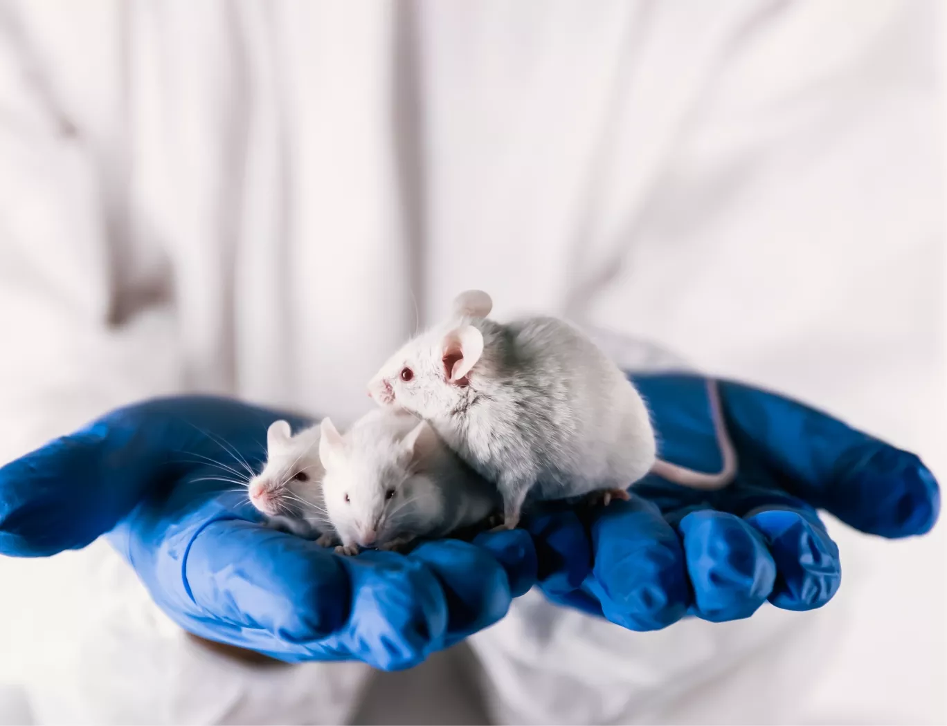
A researcher in a lab coat and blue gloves gently holds three white lab mice, symbolizing animal models in scientific research.

Quantitative Histology for LNP Formulation Selection

Traditional histology provides a qualitative assessment, which is often insufficient for the rigorous demands of comparing closely related LNP formulations. Our approach leverages automated slide scanning and advanced image analysis software to generate objective, quantitative data.

This quantitative approach delivers rapid pathology insights accelerating preclinical readouts and informing lead candidate selection. By measuring the precise percentage of tissue area occupied by lipid droplets, we provide a direct, numerical comparison of delivery efficiency between different LNP candidates. This service is supported by our >100,000 sq ft GxP facility, designed to move programs toward a successful IND submission on an 18-24 month timeline. Since 2019, programs that have engaged our full translational services have achieved a 100% IND success rate, with the Franklin Biolabs brand formally launching in 2024 to expand this offering.

Animal Welfare and GxP Compliance

All in vivo studies are conducted within AAALAC-accredited facilities under strict USDA-compliant protocols, managed through our network of qualified partners. By maximizing the data obtained from each tissue sample through quantitative digital pathology, we reduce the total number of animals required for statistically significant conclusions.

Scientific Process Diagram

This content is for informational purposes. For guidance specific to your therapeutic program, please contact our team for a consultation.