Assessing CAR-T Cell Trafficking and Persistence Using In Vivo Bioluminescence Imaging (BLI) in Solid Tumor Models

PROVEN INTELLIGENCE ACCELERATING NEXT-GENERATION THERAPIES

Assessing CAR-T Cell Trafficking and Persistence Using In Vivo Bioluminescence Imaging (BLI) in Solid Tumor Models

Assessing CAR-T Cell Trafficking and Persistence Using In Vivo Bioluminescence Imaging

CELL & GENE | RNA | BIOLOGICS

Proven Intelligence Accelerating Next-Generation Therapies.

A close-up of a pipette dispensing liquid into a rack of test tubes, set against a cool-toned, sterile background.

A scientist in protective gear pipetting a sample into a vial within a sterile laboratory hood.

Executive Summary

For CAR-T cell therapies targeting solid tumors, confirming cell trafficking to the tumor site and quantifying persistence are primary objectives of preclinical programs. Standard terminal endpoint assays like histology provide only a single snapshot in time. In vivo bioluminescence imaging (BLI) offers a quantitative, longitudinal method to repeatedly visualize and measure CAR-T cell activity within the same subject. This approach generates richer datasets to inform dose selection and minimize clinical risk, building comprehensive data packages for IND and IMPD submissions to agencies including the FDA and MHRA.

Frequently Asked Questions

    What is the primary advantage of BLI for CAR-T cell analysis?

    BLI enables longitudinal monitoring of CAR-T cell biodistribution, tumor-homing, and persistence in vivo. This provides a dynamic view of cell activity over the entire course of a study, which is not possible with terminal methods that only capture a single timepoint.

    How is the CAR-T cell signal distinguished from background noise?

    CAR-T cells are engineered to express a luciferase reporter gene. When the corresponding substrate (e.g., luciferin) is administered, only the CAR-T cells emit a light signal, which is then captured by a highly sensitive imaging system. This provides a specific and high-contrast signal.

    Can BLI data be used in regulatory submissions?

    Yes. Quantitative data from well-controlled BLI studies can be a component of the pharmacology and toxicology sections of IND and IMPD submissions. The data helps build a comprehensive picture of the therapeutic’s mechanism of action and biodistribution profile.

The Challenge of Monitoring Somatic Cell Therapies in Solid Tumors

Evaluating the in vivo behavior of CAR-T cells presents distinct challenges, particularly in solid tumor models. A therapeutic program must confirm that the engineered cells not only reach the tumor microenvironment but also expand and persist to exert their cytotoxic function.

Traditional reliance on terminal tissue collection for histology or flow cytometry provides valuable endpoint data but fails to capture the kinetics of cell trafficking and expansion. A standard, one-size-fits-all preclinical testing template does not exist for these complex therapies. A tailored, data-driven strategy is required to build a complete biological picture.

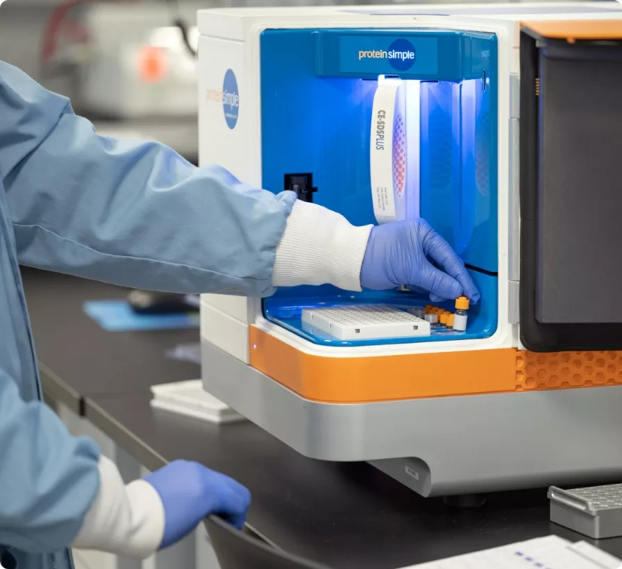
A scientist in a lab coat and gloves loads samples into a ProteinSimple instrument for analysis.

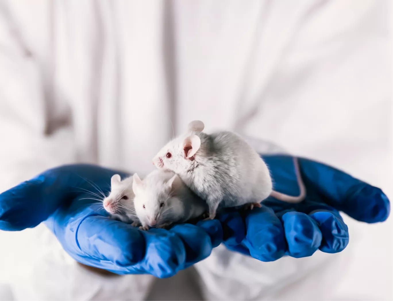
A researcher in a lab coat and blue gloves gently holds three white lab mice, symbolizing animal models in scientific research.

Longitudinal Monitoring with Bioluminescence Imaging

Bioluminescence imaging provides a noninvasive solution for tracking CAR-T cells over time. By incorporating reporter genes into the CAR-T construct, we can quantitatively visualize cell populations in vivo. This methodology allows for the direct assessment of:

  • Cell Trafficking: Confirming migration and accumulation of CAR-T cells at the primary tumor site and identifying any non-target tissue biodistribution.

  • Population Expansion: Measuring the increase in bioluminescent signal over time as an indicator of CAR-T cell proliferation in response to tumor antigens.

  • Persistence: Monitoring the duration of the CAR-T cell presence, which can offer insights into potential immune-mediated clearance mechanisms.

The ability to perform noninvasive imaging, as demonstrated in gene transfer applications (PMID: 12639305), provides a powerful translational tool. It allows for the collection of robust kinetic data from fewer animals, directly supporting a commitment to the 3Rs principles of animal research.

GxP-Compliant Programs for Regulatory Success

Data from these advanced imaging studies are integrated into comprehensive data packages designed for regulatory review. Our scientific team designs IND-enabling toxicology studies within our >100,000 sq ft facility to meet GxP expectations. While the Franklin Biolabs brand launched in 2024, our scientific leadership has maintained a 100% successful IND rate since 2019, consistently moving client candidates toward regulatory submission within an 18-24 month timeline.

A digital rendering of a DNA double helix on a dark blue background with floating particles.

A stylized, 3D rendering of a DNA double helix in light blue and white, set against a soft-focus, light gray background.

Animal Welfare and Model Selection

All studies are conducted with rigorous adherence to animal welfare guidelines established by AAALAC and the USDA. Our approach to animal care is built upon the 3Rs Principle: Replacement, Reduction, and Refinement.

Technical Visualization: In Vivo CAR-T Bioluminescence Imaging Workflow

Scientific Process Diagram

This content is for informational purposes. For guidance specific to your therapeutic program, please contact our team for a consultation.