Automated IHC Staining and Analysis for High-Throughput Preclinical Efficacy Screening

PROVEN INTELLIGENCE ACCELERATING NEXT-GENERATION THERAPIES

Automated IHC Staining and Analysis for High-Throughput Preclinical Efficacy Screening

CELL & GENE | RNA | BIOLOGICS
  • Precision Histology at Scale: Quantitative, Reproducible Biomarker Assessment.*

Franklin Biolabs’ automated immunohistochemistry (IHC) and quantitative analysis platform provides high-throughput, objective biomarker assessment for preclinical efficacy and non-target tissue biodistribution studies. By replacing subjective manual scoring with algorithm-driven analysis, our platform generates reproducible, cell-by-cell data to support accelerated program decisions and strengthen regulatory submissions.

    What platforms do you use for automated IHC?

    Our GxP-compliant histology laboratory is equipped with Leica BOND RX systems for fully automated IHC and ISH staining, ensuring high reproducibility and run-to-run consistency for large-scale studies.

    How does automated analysis differ from manual pathologist scoring?

    Automated analysis provides objective, quantitative data by removing inter- and intra-observer variability. Instead of semi-quantitative scores, our platform delivers precise metrics such as positive cell counts, H-scores, and spatial distribution of biomarkers on a cell-by-cell basis across entire tissue sections.

    What is the typical turnaround time for a high-throughput IHC study?

    Timelines are project-dependent and defined by study scale. However, the combination of automated staining and high-speed whole-slide scanning significantly reduces the time required compared to manual methods, enabling faster data delivery for key program milestones.

    Can you develop custom IHC assays for novel targets?

    Yes. Our scientific team specializes in rigorous antibody validation and protocol optimization for novel targets. We establish robust, specific, and reproducible assays qualified for use in GxP-compliant preclinical studies.

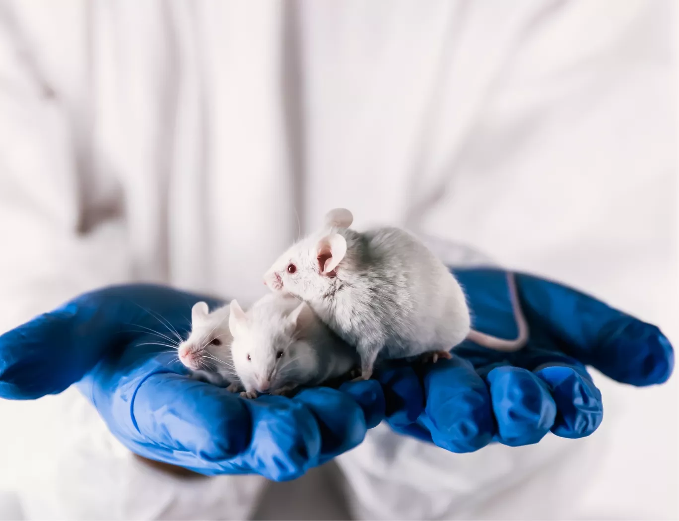
A researcher in a lab coat and blue gloves gently holds three white lab mice, symbolizing animal models in scientific research.

The Challenge of Scalable and Objective Histology

In preclinical development, particularly for cell and gene therapies, evaluating efficacy and non-target tissue biodistribution requires analyzing a substantial volume of tissue samples. Traditional manual histology methods, reliant on pathologist scoring, introduce variability and are not scalable for the demands of modern therapeutic programs. This subjectivity can obscure subtle but meaningful biological effects, complicating data interpretation and delaying decisions.

A Quantitative Approach to Biomarker Assessment

Franklin Biolabs addresses this challenge with a fully automated workflow for IHC staining and quantitative image analysis. Our process standardizes every step from slide preparation to data extraction, delivering objective and reproducible biomarker quantification. This commitment to precision is analogous to advancements in molecular biology. For instance, the use of high-fidelity polymerases is recognized as fundamental for the accurate amplification and discovery of novel AAV variants from tissue (PMID: 34448594). The principle is the same: the integrity of the final data is wholly dependent on the fidelity of the methods used to generate it.

Our platform provides:

  • Automated Staining: Reduces technical variability for consistent results across large sample cohorts.

  • Whole-Slide Digital Scanning: Creates high-resolution digital archives for permanent records and re-analysis.

  • AI-Powered Image Analysis: Applies validated algorithms to detect, segment, and quantify biomarkers objectively.

This systematic approach, conducted within our >100,000 sq ft facility, provides the Rapid Pathology Insights Accelerating Preclinical Readouts needed to advance complex programs.

A digital rendering of a DNA double helix on a dark blue background with floating particles.

A stylized, 3D rendering of a DNA double helix in light blue and white, set against a soft-focus, light gray background.

Platform Capabilities Comparison

Parameter Manual Histology Automated IHC & Quantitative Analysis
Throughput Low to Moderate High
Objectivity Subjective (Observer-Dependent) Objective (Algorithm-Driven)
Data Type Semi-Quantitative (0, 1+, 2+, 3+) Quantitative (Cell Counts, H-Score)
Reproducibility Moderate High
Data Granularity Regional Estimation Single-Cell Resolution

Accelerating Your IND Timeline

Robust, quantitative, and reproducible pathology data is a cornerstone of a successful Investigational New Drug (IND) application. Our automated platform generates the high-quality datasets needed to build a compelling narrative around your asset’s mechanism of action and safety profile. This level of documentation supports an accelerated path toward regulatory submission, aligning with our typical 18-24 month IND timeline. Since the Franklin Biolabs brand launch in 2024, programs leveraging our comprehensive preclinical services have maintained a 100% IND success rate, a record established by our core scientific team since 2019.

Scientific Process Diagram

This content is for informational purposes. For guidance specific to your therapeutic program, please contact our team for a consultation.