Biodistribution and Persistence of Lipid Nanoparticles Following Intramuscular Administration

PROVEN INTELLIGENCE ACCELERATING NEXT-GENERATION THERAPIES

Biodistribution and Persistence of Lipid Nanoparticles Following Intramuscular Administration

CELL & GENE | RNA | BIOLOGICS
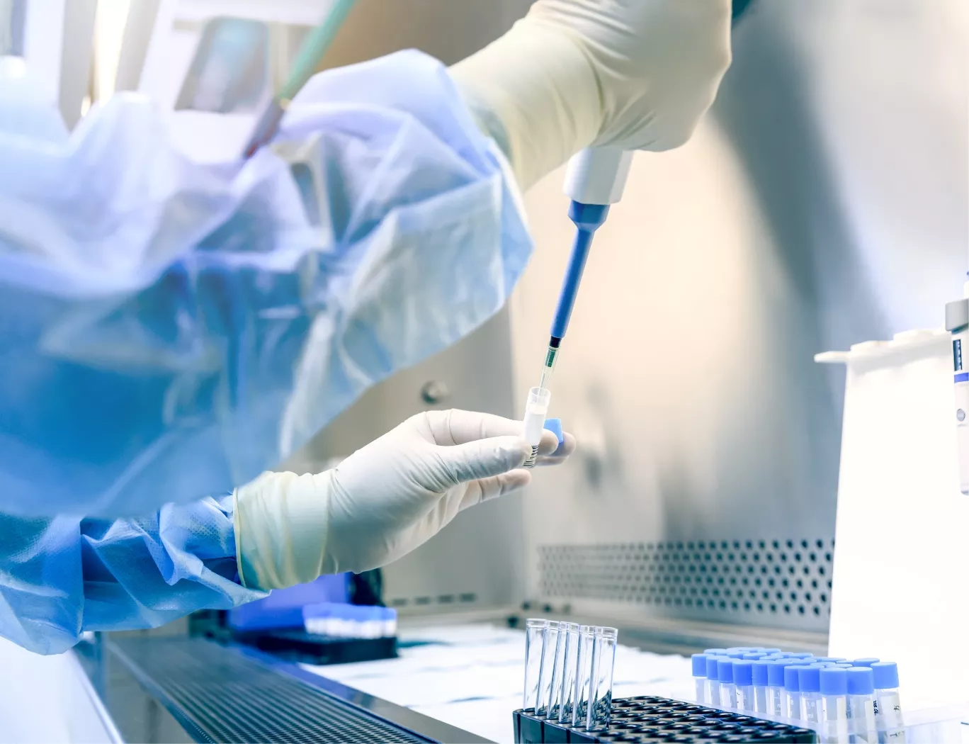
A scientist in protective gear pipetting a sample into a vial within a sterile laboratory hood.

Executive Summary

The biodistribution of lipid nanoparticles (LNPs) following intramuscular administration is not uniform; it is dictated by specific formulation components. Helper lipids, for instance, directly modulate protein corona formation, which in turn governs organ tropism, primarily to the liver and spleen. A comprehensive understanding and quantification of this distribution and persistence profile is a prerequisite for building a robust safety package to support Investigational New Drug (IND) applications.

Frequently Asked Questions

    What are the primary factors influencing LNP biodistribution after intramuscular injection?

    A: Key determinants include the LNP formulation itself (e.g., helper lipids, ionizable lipids), particle size, surface charge, and the subsequent formation of a protein corona upon entering the systemic circulation.

    How do you quantify LNP persistence in tissues?

    A: We employ a multi-modal approach combining quantitative methods, such as qPCR for mRNA or siRNA payloads and LC-MS/MS for lipid components, with qualitative and semi-quantitative Histology to confirm tissue localization and assess any associated pathological findings.

    Which tissues are most relevant for non-target tissue biodistribution analysis of LNPs?

    A: The liver and spleen are well-established primary sites of LNP accumulation. Our study designs also include rigorous evaluation of the injection site, draining lymph nodes, and other key organs as defined by regulatory guidance to build a complete safety profile.

The Influence of Formulation on LNP Trafficking

The selection of helper lipids is a primary determinant of LNP organ tropism. This represents an active biological interaction driven by the specific protein corona that forms around the nanoparticle surface upon systemic exposure.

Recent investigations have clarified how different helper lipids alter this protein adsorption profile, directly influencing biodistribution. As demonstrated in high-throughput in vivo screening (PMID: 33404020), formulations containing certain helper lipids like DOPE exhibit high apolipoprotein E (ApoE) adsorption. This interaction preferentially directs the LNPs to the liver. Conversely, formulations using lipids such as DSPC result in lower ApoE binding, leading to increased accumulation in the spleen.

This has direct consequences for therapeutic development:

  • Efficacy: The choice of helper lipid can be engineered to enhance payload delivery to a desired organ system.

  • Safety: Understanding this predictable distribution allows for a more focused assessment of potential on-target or non-target tissue toxicities.

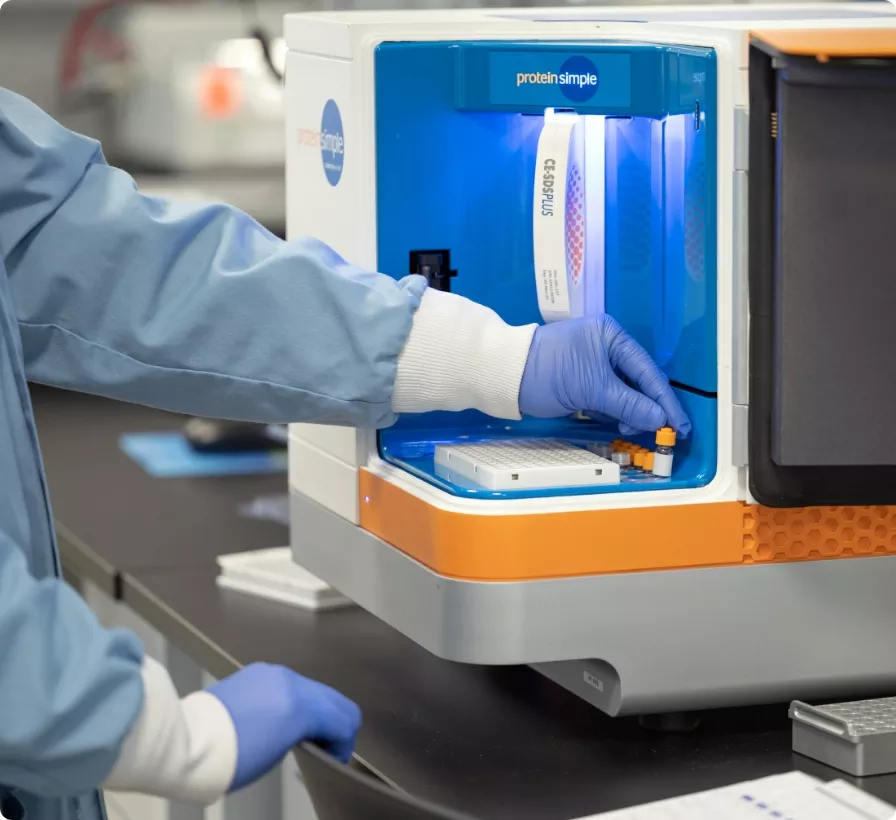
A scientist in a lab coat and gloves loads samples into a ProteinSimple instrument for analysis.

A researcher in a lab coat and blue gloves gently holds three white lab mice, symbolizing animal models in scientific research.

Quantifying Distribution for a Regulatory-Grade Safety Profile

Translating these formulation insights into a successful regulatory submission requires precise, GxP-compliant quantification. Our programs integrate bioanalytical assays with comprehensive Histology to map the kinetics of LNP distribution and persistence. This detailed characterization provides the robust safety data necessary to support clinical translation.

At our >100,000 sq ft facility, we execute these complex studies to support an average 18-24 month IND timeline for our sponsors. Since 2019, programs we have supported have achieved a 100% IND success rate, a record of quality we continue with the Franklin Biolabs brand, which launched in 2024.

Animal Welfare and Study Design

All in vivo studies are designed to maximize the scientific value obtained while adhering to the highest ethical standards for animal welfare. Our programs, accredited by AAALAC and regulated by the USDA, are structured around the 3Rs principles (Replacement, Reduction, and Refinement). By implementing advanced bioanalytical techniques and multiplexed tissue analysis, we reduce the total number of animals required for a complete biodistribution and toxicology program.

Scientific Process Diagram

This content is for informational purposes. For guidance specific to your therapeutic program, please contact our team for a consultation.