IHC for Assessing Target Engagement of Bi-specific T-cell Engagers (BiTEs) in Tumor Tissues

PROVEN INTELLIGENCE ACCELERATING NEXT-GENERATION THERAPIES

IHC for Assessing Target Engagement of Bi-specific T-cell Engagers (BiTEs) in Tumor Tissues

Immunohistochemistry (IHC) for Target Engagement of Bi-specific T-cell Engagers (BiTEs)

CELL & GENE | RNA | BIOLOGICS

Visualizing Mechanism of Action at Cellular Resolution

The efficacy of a Bi-specific T-cell Engager (BiTE) is contingent upon its ability to physically bridge a T-cell and a tumor cell within the tumor microenvironment (TME). While systemic assays can measure pharmacodynamics, they cannot confirm this spatial co-localization. Immunohistochemistry (IHC) provides definitive, tissue-based evidence of target engagement, visualizing the induced immune synapse directly within tumor samples. This analysis is a key component of the data package required to demonstrate mechanism of action (MOA) for IND-enabling studies.

What is the primary application of IHC for BiTE analysis?
To visually confirm the mechanism of action by demonstrating T-cell (e.g., CD3+) infiltration and direct proximity to tumor cells expressing the target antigen within the complex architecture of the TME.
Which markers are typically used in a BiTE IHC panel?
A standard panel includes a pan T-cell marker (CD3), a tumor-specific antigen (e.g., CD20, BCMA), and can be expanded to include proliferation markers (Ki-67) or immune checkpoint proteins (PD-1, CTLA-4) to assess the functional state of the engaged T-cells.
How does multiplex IHC (mIHC) enhance BiTE assessment?
Multiplex IHC allows for the simultaneous visualization of multiple cellular phenotypes and functional states on a single tissue section. This provides deep spatial context, enabling the characterization of complex cellular neighborhoods and the interplay between T-cell activation, tumor cell killing, and potential resistance mechanisms.
What are the GxP considerations for IHC assays supporting regulatory submissions?
Assays intended for regulatory filings must be developed and validated under a GxP framework. This process ensures the antibody reagents and staining protocols are specific, sensitive, and highly reproducible, guaranteeing the integrity and reliability of the data submitted in an IND package.

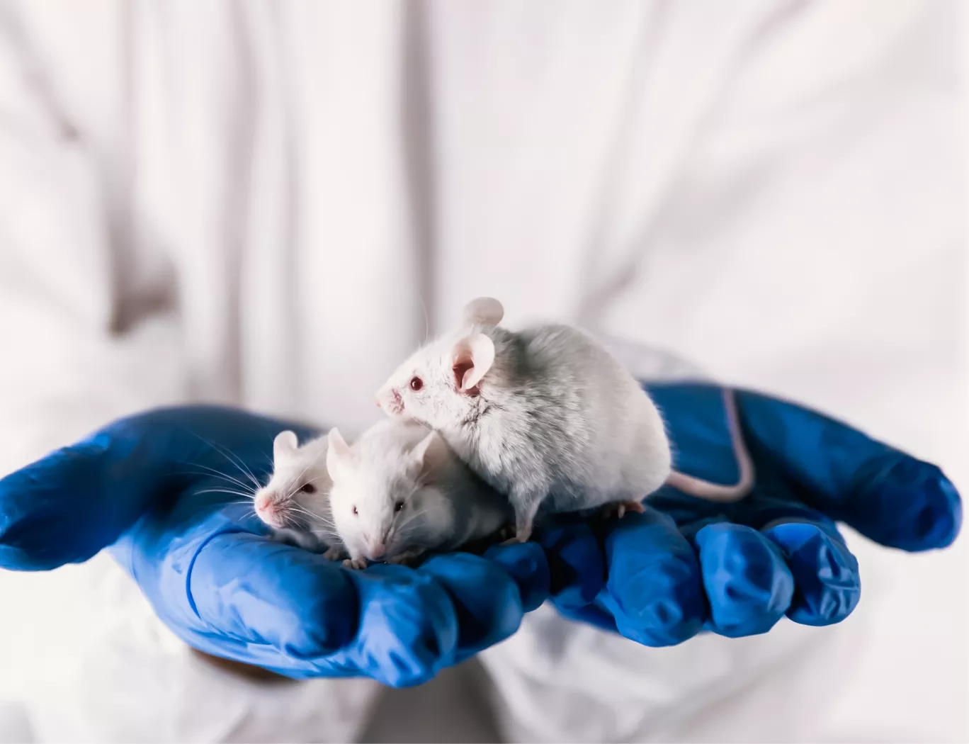
A researcher in a lab coat and blue gloves gently holds three white lab mice, symbolizing animal models in scientific research.

The Challenge of Quantifying BiTE Mechanism of Action in Tissue

Bi-specific T-cell Engagers function by forcing an interaction between cytotoxic T-cells and malignant cells. Verifying that this engineered synapse occurs as intended within solid tumors or hematological tissues is a primary objective of preclinical assessment. Systemic readouts from peripheral blood are insufficient, as they do not reflect the biological activity occurring within the target tissue.

IHC and immunofluorescence (IF) are the gold-standard methods for providing direct visual proof of this MOA. By using specific antibodies against T-cell and tumor antigens, these techniques map the precise location of each cell type, confirming that the therapeutic has successfully recruited immune effectors to the site of disease. This approach provides the Rapid Pathology Insights Accelerating Preclinical Readouts necessary to build confidence in a candidate molecule before moving forward.

Advanced Preclinical Imaging and Analysis

The infrastructure required to execute these complex assays at scale is a significant consideration in program planning. Franklin Biolabs supports these studies with dedicated Histology and digital pathology platforms.

A digital rendering of a DNA double helix on a dark blue background with floating particles.

A stylized, 3D rendering of a DNA double helix in light blue and white, set against a soft-focus, light gray background.

Parallels in T-cell Response Characterization

The need to accurately characterize T-cell responses in tissue is a shared challenge across many advanced therapeutic platforms. For example, investigations into AAV vector biology identified the specific capsid epitopes responsible for eliciting CD8+ T-cell responses, providing tools to monitor cellular immunity (PMID: 19777488).

Subsequent work demonstrated that such transgene-specific T-cell responses can have profound functional consequences, including the potential for tissue damage and loss of therapeutic protein expression in preclinical models (PMID: 19441963). This body of research highlights a unifying principle: whether mitigating an unwanted immune response in gene therapy or driving a desired one with a BiTE, robust, tissue-based analytical methods are fundamental to understanding and predicting biological outcomes.

Franklin Biolabs GxP Histology Capabilities

Our team provides comprehensive Histology services within our >100,000 sq ft facility to support programs from discovery through IND submission. We develop and validate custom IHC assays for novel targets, leveraging our digital pathology platforms for quantitative, objective analysis. This rigorous, data-driven approach helps de-risk therapeutic programs, supporting an 18-24 month IND timeline. Since 2019, programs we have supported have maintained a 100% IND success rate; the Franklin Biolabs brand itself launched in 2024.

The table below outlines two common approaches for BiTE analysis.

Feature Standard Dual-Label IHC Multiplex IHC (mIHC)
Markers per Section 2-3 4-8+
Spatial Context Good (Proximity of two cell types) Excellent (Complex cellular neighborhoods)
Data Output Semi-quantitative scores, cell counts High-plex spatial data, cell phenotype mapping
Application Confirmation of MOA (T-cell:Tumor) Deep TME characterization, resistance mechanisms

Animal Welfare and Programmatic Alignment

Our commitment to the 3Rs (Reduction, Refinement, Replacement) is a core tenet of our scientific operations, fully accredited by AAALAC and compliant with USDA regulations. By employing advanced multiplexing techniques that maximize the data derived from each tissue sample, we directly support the principle of Reduction, obtaining deeper biological insights while minimizing the number of animals required for a study.

Scientific Process Diagram

This content is for informational purposes. For guidance specific to your therapeutic program, please contact our team for a consultation.