Integrating Whole Slide Imaging and AI for Predictive Toxicology in Preclinical Biologic Development

PROVEN INTELLIGENCE ACCELERATING NEXT-GENERATION THERAPIES

Integrating Whole Slide Imaging and AI for Predictive Toxicology in Preclinical Biologic Development

CELL & GENE | RNA | BIOLOGICS

Frequently Asked Questions (FAQ)

    What is Whole Slide Imaging (WSI)?

    A: Whole Slide Imaging is a digital pathology technology that uses automated microscopy to capture an entire histological slide at high magnification. This process creates a single, high-resolution digital file, or “virtual slide,” enabling remote viewing, sharing, and computational analysis without a physical microscope.

    How does AI enhance traditional histology for biologics?

    A: AI algorithms applied to WSI data provide objective, quantitative analysis at a scale unachievable with manual pathology. For biologics, AI can precisely quantify cellular responses, measure vector biodistribution, and detect subtle morphometric changes across entire tissue sections, replacing subjective scoring with reproducible, statistically robust data for IND-enabling studies.

    What WSI data formats does Franklin Biolabs support?

    A: Our platform is agnostic and supports all major proprietary WSI formats (e.g., .svs, .ndpi, .czi) as well as the DICOM standard for medical imaging. We provide sponsors with both the raw image files and the quantitative analysis output.

    How does this approach de-risk an IND submission?

    A: By generating quantitative, objective toxicology and biodistribution data, this method strengthens the regulatory package. It minimizes the inter-observer variability common in manual pathology reads, providing a more defensible and reproducible safety profile to regulatory agencies. This clarity supports our typical 18-24 month IND timeline.

Executive Summary

The integration of high-throughput Whole Slide Imaging (WSI) with artificial intelligence (AI) provides a quantitative, scalable solution for preclinical toxicology assessment. This computational pathology approach moves beyond traditional semi-quantitative scoring to deliver objective, reproducible data on cellular morphology, protein expression, and non-target tissue biodistribution. For sponsors developing complex biologics, this methodology generates a more robust and defensible dataset to support accelerated Investigational New Drug (IND) application timelines.

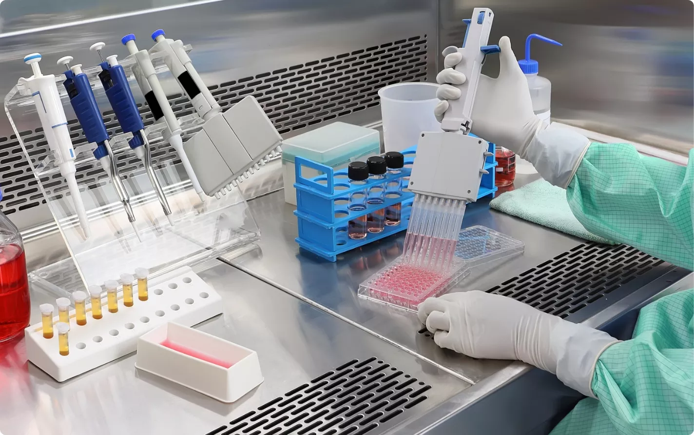
A scientist in a sterile laboratory setting uses a multichannel pipette to transfer pink liquid into a multi-well plate for a high-throughput experiment.

The Scaling Challenge in Preclinical Histology

Standard histology workflows rely on manual microscopic evaluation by a board-certified pathologist. While effective, this method presents scalability and objectivity challenges when applied to the large, multi-tissue studies required for biologic safety assessment. Semi-quantitative scoring can introduce variability, and detecting subtle, diffuse toxicological signals across thousands of slides is a significant logistical undertaking. This is particularly true for novel constructs like cell therapies or RNA-based platforms, where the mechanism of toxicity may be nuanced.

A Quantitative Framework: WSI and AI Deployment

Our GxP-compliant histology services, conducted within our >100,000 sq ft facility, leverage a fully digitized workflow.

  • High-Throughput Digitization: Tissue sections are processed and then scanned to create high-resolution virtual slides.

  • Algorithm-Driven Analysis: We deploy validated AI models to perform specific analytical tasks directly on these digital images. This includes:

    • Automated cell counting and classification.

    • Morphometric analysis of cellular and tissue structures.

    • Quantification of biomarker expression (IHC/ISH).

    • Mapping of non-target tissue biodistribution for novel vectors.

  • Objective Data Output: The system generates quantitative data, removing the subjectivity of manual scoring. This provides clear, statistically significant results that directly inform safety and efficacy assessments, yielding rapid pathology insights that accelerate preclinical readouts.

This level of precision in evaluation is the necessary counterpart to advancements in payload delivery. For example, techniques enabling MRI-guided stereotactic injections are setting new standards for targeting accuracy (PMID: 38310346). A computational pathology platform is required to verify and quantify the on-target efficacy and off-target safety profile resulting from such precise administration. The core scientific team, now leading Franklin Biolabs following the brand’s 2024 launch, has maintained a 100% IND success rate for client programs since 2019.

A blue-toned image of white lab rats in their cages within a laboratory or vivarium setting, likely for scientific research or testing.

Commitment to Enhanced Animal Welfare

Our approach to computational pathology directly supports the 3Rs principles of animal welfare (Replacement, Reduction, and Refinement). This commitment is reflected in programs conducted to the standards required for AAALAC accreditation and USDA oversight. By extracting maximal quantitative data from every tissue sample, we refine study outcomes and can often reduce the number of animals required to achieve statistical power.

Scientific Process Diagram

This content is for informational purposes. For guidance specific to your therapeutic program, please contact our team for a consultation.