Multi-Modal In Vivo Imaging (MRI, PET, BLI) for Comprehensive Pharmacodynamic Assessment of Cell Therapies

PROVEN INTELLIGENCE ACCELERATING NEXT-GENERATION THERAPIES

Multi-Modal In Vivo Imaging (MRI, PET, BLI) for Comprehensive Pharmacodynamic Assessment of Cell Therapies

Multi-Modal In Vivo Imaging for Cell Therapy Pharmacodynamics

CELL & GENE | RNA | BIOLOGICS
Proven Intelligence Accelerating Next-Generation Therapies.

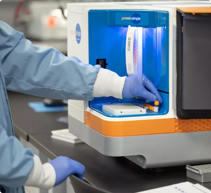
A scientist in a lab coat and gloves loads samples into a ProteinSimple instrument for analysis.

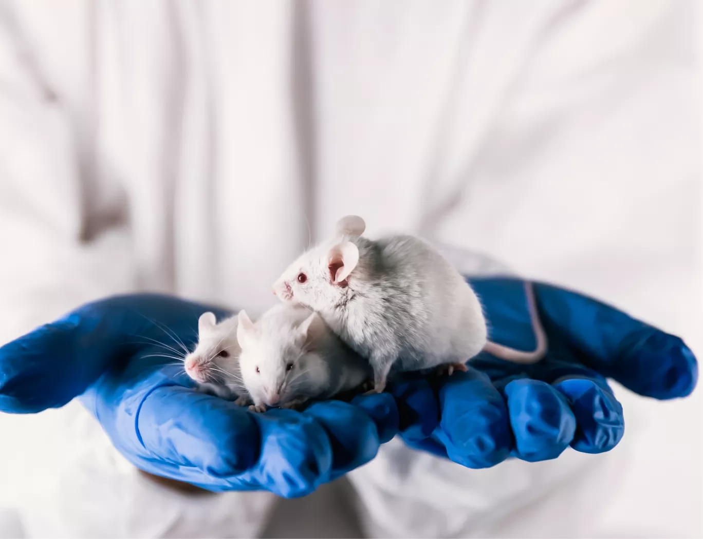
A researcher in a lab coat and blue gloves gently holds three white lab mice, symbolizing animal models in scientific research.

Executive Summary

Traditional pharmacodynamic (PD) assessments that rely on terminal endpoints are insufficient for characterizing the complex, dynamic behavior of cell-based therapeutics like CAR-T, CAR-NK, or iPSC-derived candidates. This page details a multi-modal in vivo imaging strategy that combines Magnetic Resonance Imaging (MRI), Positron Emission Tomography (PET), and Bioluminescence Imaging (BLI) to generate a longitudinal, quantitative data package. This approach provides a comprehensive understanding of cell trafficking, persistence, and functional activity, minimizing clinical risk and supporting a robust IND submission within an 18-24 month timeline.

Frequently Asked Questions

Why is multi-modal imaging necessary for cell therapies?

Cell therapies are living therapeutics whose efficacy depends on their ability to traffic to a target site, expand, persist, and exert a functional effect. A single imaging modality provides an incomplete picture. BLI offers high-sensitivity tracking of cell viability, PET can quantify metabolic activity or receptor engagement, and MRI provides high-resolution anatomical context. Combining them creates a comprehensive, longitudinal view of the therapeutic’s in vivo behavior.

How does this imaging strategy support IND-enabling toxicology studies?

By non-invasively monitoring cell biodistribution in real-time, we can identify potential accumulation in non-target tissues early in development. This provides substantive data for GxP-compliant toxicology programs, informing dose selection and safety assessments without relying exclusively on terminal histology. This approach strengthens the regulatory submission by demonstrating a deep understanding of the therapeutic’s in vivo fate.

Can these imaging techniques be applied in NHP models?

Yes. Our >100,000 sq ft facility is equipped for advanced imaging in both rodent and nonhuman primate models. The principles of MRI-guided administration and PET/SPECT-based quantitative imaging are directly translatable to NHP models, providing pivotal data on the pharmacodynamics and safety profile in a higher-order species before human trials.

The Limitations of Terminal Endpoints

Assessing the in vivo activity of somatic cell therapies presents a distinct set of challenges. Unlike static biologics, the biodistribution and functional persistence of CAR-T, TCR-T, or stem cell therapeutics are dynamic processes. Relying solely on terminal histology and tissue analysis provides only a single snapshot in time, failing to capture the kinetics of cell trafficking, expansion, and clearance.

A tailored preclinical strategy is required to build a complete narrative of the therapeutic’s mechanism of action. This involves generating longitudinal data that visualizes and quantifies the asset’s behavior over the full course of a response.

A digital rendering of a DNA double helix on a dark blue background with floating particles.

A stylized, 3D rendering of a DNA double helix in light blue and white, set against a soft-focus, light gray background.

A Multi-Modal Imaging Strategy

A more robust approach integrates several non-invasive imaging modalities to build a complete pharmacodynamic profile. Each technique provides a unique layer of information, creating a comprehensive dataset.

  • Bioluminescence Imaging (BLI): Ideal for high-throughput, sensitive tracking of luciferase-expressing cells. BLI provides a clear signal for cell localization and viability over time.

  • Positron Emission Tomography (PET): Offers quantitative data on metabolic activity or receptor occupancy. Using specific radiotracers, PET can confirm not just where the cells are, but if they are functionally active.

  • Magnetic Resonance Imaging (MRI): Delivers high-resolution anatomical detail. MRI is used to co-register functional data from BLI or PET, providing precise anatomical context for cell localization and assessment of downstream physiological effects, such as changes in tumor volume.

This combination of techniques generates High-Fidelity In Vivo Intelligence for Complex Modalities.

Precision Delivery and Quantitative Monitoring

The quality of a pharmacodynamic assessment begins with reproducible administration. As demonstrated in advanced vector applications, MRI-guided frameless stereotactic systems enable highly accurate, real-time visualization of therapeutic delivery to specific anatomical locations (PMID: 38310346). This technical precision minimizes variability between subjects and ensures that observed effects are a direct result of the therapeutic’s action at the intended site.

Following administration, quantitative monitoring of the therapeutic is a requirement for a robust data package. The principles established for non-invasive imaging of gene transfer, using reporter systems to track payload expression via imaging like SPECT, can be extrapolated to cell therapies (PMID: 12639305). This allows for longitudinal quantification of cell persistence and activity, providing a powerful dataset for modeling human dose-response relationships.

A scientist in a sterile laboratory setting uses a multichannel pipette to transfer pink liquid into a multi-well plate for a high-throughput experiment.

Animal Welfare and Enhanced Data Collection

Implementing a longitudinal, non-invasive imaging strategy directly aligns with our commitment to the 3Rs principle (Replacement, Reduction, and Refinement) of animal welfare. By collecting multiple data points from the same animal over time, this approach significantly reduces the total number of animals required for a study compared to designs that depend on separate cohorts for each terminal timepoint.

Our programs are AAALAC accredited and USDA compliant, with a focus on enhanced housing and social enrichment.

Technical Visualization: In Vivo Multi-Modal Imaging Workflow

Scientific Process Diagram

This content is for informational purposes. For guidance specific to your therapeutic program, please contact our team for a consultation.