Pathology Support for Preclinical Safety Assessment of Engineered NK Cell Therapies

PROVEN INTELLIGENCE ACCELERATING NEXT-GENERATION THERAPIES

Pathology Support for Preclinical Safety Assessment of Engineered NK Cell Therapies

CELL & GENE | RNA | BIOLOGICS

A robust preclinical pathology program is fundamental to de-risking engineered Natural Killer (NK) cell therapies. The primary objective is to characterize cellular biodistribution, persistence, and any potential on-target, off-tumor or non-target tissue toxicities. This requires a specialized histology approach that moves beyond standard assessments to incorporate multiplexed biomarker analysis and quantitative digital pathology, ensuring a clear, data-driven path toward a successful Investigational New Drug (IND) application.

Technical Question Franklin Biolabs Approach
What are the primary pathology endpoints for an engineered NK cell safety study? Key endpoints include quantifying non-target tissue biodistribution, evaluating potential on-target/off-tumor activity, monitoring for signs of cytokine release syndrome (CRS) at the tissue level, and assessing NK cell persistence or engraftment using highly specific markers.
How do you differentiate therapeutic effect from off-target toxicity in tissue samples? We utilize advanced multiplex immunofluorescence (mIF) and immunohistochemistry (mIHC) to visualize the NK cell, its target antigen, and markers of cell death simultaneously. This co-localization data, combined with quantitative image analysis, provides definitive evidence to distinguish intended cytotoxicity from unintended effects.
What GxP standards apply to preclinical pathology for cell therapies? All study phases, from necropsy and tissue processing through slide analysis and reporting, are conducted within a rigorous GxP framework. This ensures the data integrity, traceability, and quality required for regulatory submissions to agencies like the MHRA and FDA.

A 3D rendering of Y-shaped antibody molecules against a blue, abstract background.

A close-up of a scientist in a lab, wearing blue gloves and examining the results of a gel electrophoresis or Western blot.

Defining the Safety Profile of Engineered NK Cells

The unique biological characteristics of engineered NK cells, including their potential for allogeneic application and distinct cytotoxicity mechanisms, demand a bespoke pathology evaluation. A comprehensive safety assessment must accurately characterize the kinetics and activity of these therapies in vivo. This involves meticulously evaluating tissues for evidence of unintended cellular damage or immune activation away from the tumor site.

Our approach focuses on several key areas:

  • Cellular Biodistribution: Determining where the engineered NK cells traffic and accumulate.

  • On-Target, Off-Tumor Evaluation: Assessing activity in healthy tissues that may express the target antigen at low levels.

  • Host Immune Response: Characterizing any local immune cell infiltration or reaction to the therapeutic cells.

  • Cell Persistence: Using specific markers to track the longevity of the NK cell population post-administration.

A Multi-Modal Approach to Histology and Analysis

Standard hematoxylin and eosin (H&E) staining provides a necessary baseline, but a deeper understanding requires more advanced techniques. We deploy a suite of technologies to build a complete picture of the engineered NK cell’s behavior within the tissue microenvironment. This quantitative approach provides objective data, enabling confident and timely decision-making.

Our analytical toolkit includes:

  • Multiplex Immunohistochemistry (mIHC/mIF): To simultaneously identify NK cells, target cells, and various immune infiltrates (e.g., T-cells, macrophages) within a single tissue section.

  • In Situ Hybridization (ISH): To detect specific genetic modifications or payloads, confirming the identity and location of the engineered cells.

  • Digital Pathology & AI-Powered Image Analysis: We leverage our strategic partnership with industry leaders to apply algorithms for objective, quantitative assessment of cell distribution, proliferation, and tissue architecture changes.

A close-up of a pipette dispensing liquid into a rack of test tubes, set against a cool-toned, sterile background.

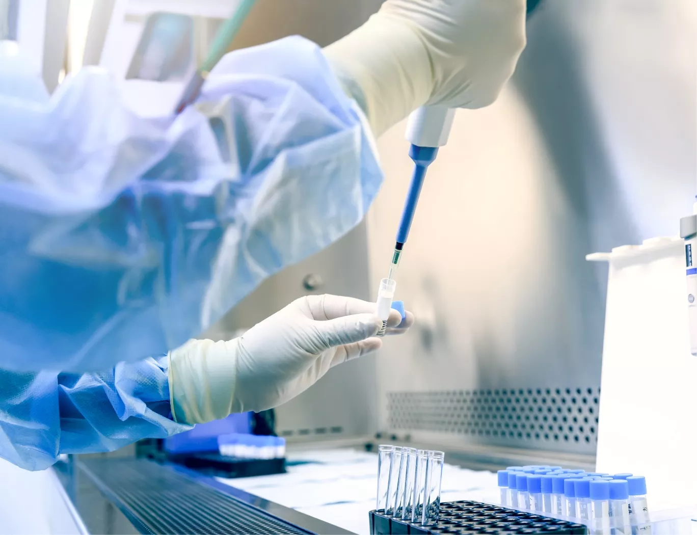
A scientist in protective gear pipetting a sample into a vial within a sterile laboratory hood.

Integrating Pathology with Broader Safety Endpoints

Histology data does not exist in a vacuum. Its value is maximized when correlated with other study endpoints like clinical chemistry and systemic cytokine profiling. For example, observations of immune cell infiltration in non-target tissues can be mechanistically linked to specific cytokine signatures, helping to differentiate systemic inflammatory responses from direct cell-mediated effects. This integrated analysis, combining quantitative pathology with systemic biomarker data, is instrumental in building a comprehensive and predictive safety model. By integrating our findings, we provide a cohesive narrative that supports the overall safety profile of the therapeutic candidate.

Animal Welfare and GxP Compliance

Our >100,000 sq ft GxP-compliant facility is designed to support complex cell therapy programs, helping sponsors navigate the typical 18-24 month IND timeline. While the Franklin Biolabs brand was established in 2024, our core scientific team has maintained a 100% IND-enabling study success rate since 2019.

Scientific Process Diagram

This content is for informational purposes. For guidance specific to your therapeutic program, please contact our team for a consultation.