Quantitative Digital Pathology for AAV9 Biodistribution in CNS Tissues for Boston Biotech

PROVEN INTELLIGENCE ACCELERATING NEXT-GENERATION THERAPIES

Quantitative Digital Pathology for AAV9 Biodistribution in CNS Tissues for Boston Biotech

Quantitative Digital Pathology for AAV9 Biodistribution in CNS Tissues

CELL & GENE | RNA | BIOLOGICS

Frequently Asked Questions (FAQ)

Technical Question Franklin Biolabs Response
What is the standard resolution for whole-slide imaging of CNS tissue? Our platforms operate at a 40x objective magnification, enabling cellular-level resolution for precise quantification of vector localization and transgene expression within complex neuroanatomical structures.
How do you differentiate vector genetic material from transduced cell expression? We employ a multi-modal approach. In situ hybridization (ISH) is used to detect vector genetic material, while immunohistochemistry (IHC) targets the expressed transgene protein. Co-localization analysis on serial sections provides a clear distinction.
What controls validate AAV9 capsid staining in brain tissue? We utilize a robust control strategy including: 1) tissue from naive, untreated animals as a negative control, 2) isotype control antibodies to rule out non-specific binding, and 3) a well-characterized positive control AAV9 vector.
Can your platform quantify biodistribution in specific brain regions? Yes. Our digital pathology software, supported by our board-certified pathologists, allows for precise annotation of specific neuroanatomical regions (e.g., hippocampus, cerebellum, cortex) for region-specific quantitative analysis.

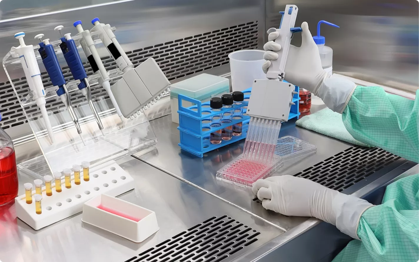
A scientist in a sterile laboratory setting uses a multichannel pipette to transfer pink liquid into a multi-well plate for a high-throughput experiment.

Executive Summary

For biotechnology companies developing AAV9-based gene therapies for central nervous system (CNS) disorders, accurately quantifying vector biodistribution is a primary determinant of a successful regulatory submission. Intrathecal administration does not guarantee CNS confinement; a significant portion of the vector dose can distribute systemically. Franklin Biolabs utilizes quantitative digital pathology and AI-driven image analysis to provide objective, cellular-level biodistribution data for both on-target CNS regions and non-target peripheral tissues. This approach generates the robust, reproducible datasets required to support safety assessments and accelerate the typical 18-24 month timeline to an Investigational New Drug (IND) application.

The Challenge of Systemic Exposure After CNS Dosing

Direct administration of AAV vectors to the cerebrospinal fluid (CSF) is a common strategy for targeting neurological disease. However, this route does not prevent vector entry into systemic circulation. As demonstrated in nonclinical studies, a substantial fraction of the administered dose can escape the CNS and distribute to peripheral organs (PMID: 37624734). This non-target tissue biodistribution has direct implications for:

  • Dose-Limiting Toxicity: Unintended vector accumulation in organs like the liver or dorsal root ganglia can lead to adverse findings.

  • Immunogenicity: Systemic exposure can trigger an immune response against the AAV capsid, potentially impacting efficacy and safety.

  • Translational Modeling: Accurately defining the relationship between dose, CNS exposure, and systemic exposure is fundamental for predicting a safe and effective human starting dose.

A blue-toned image of white lab rats in their cages within a laboratory or vivarium setting, likely for scientific research or testing.

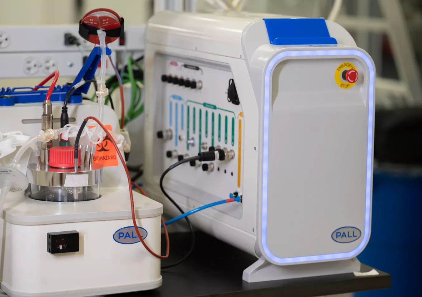
A close-up shot of a modern bioreactor system and control unit from Pall Corporation in a clean laboratory environment.

A Quantitative Approach to AAV9 Biodistribution

Qualitative assessments from traditional histology are no longer sufficient for building a comprehensive IND package. We provide a quantitative framework to de-risk AAV9 biodistribution studies. Our pathology services combine automated staining with high-throughput whole-slide scanning and algorithm-based image analysis to deliver objective data.

This platform enables:

  • Cellular-Level Quantification: Measuring the percentage of positive cells and staining intensity within specific, pathologist-annotated regions.

  • Objective Data Outputs: Generating reproducible numerical data instead of subjective scoring, strengthening the regulatory submission.

  • Comprehensive Tissue Evaluation: Analyzing both CNS and a full panel of peripheral tissues under GxP conditions in our >100,000 sq ft facility.

These methods provide the clear, defensible data packages that support an accelerated development path. Franklin Biolabs has maintained a 100% IND-enabling program success rate since 2019, with the Franklin Biolabs brand itself launching in 2024 to continue this record of excellence.

)

Integrating Data for a Stronger Regulatory Position

The output from our quantitative pathology platform integrates directly with other key preclinical endpoints, such as qPCR/ddPCR for vector copy number and bioanalytical data for transgene expression. This multi-faceted approach builds a cohesive and convincing narrative for regulatory review, providing rapid pathology insights that accelerate preclinical readouts.

“We started collaborating with UPenn Vector core in 2023 and the AAV vector which they manufactured laid a foundation for development of a gene therapy candidate which will enter soon preclinical studies. The key people from UPenn Vector Core joined Franklin Biolabs and our partnership transitioned without interruption from UPenn Vecor Core to Franklin Biolabs Research Vector Division. Franklin Biolabs is our trusted partner in our AAV-vector based gene therapy candidate development and we hope to continue the partnership for years to come.”
— Biotech Partner

A stylized 3D rendering of a DNA double helix, composed of light-colored spheres on a translucent blue backbone, set against a soft-focus, light blue background.

Commitment to Animal Welfare

All in vivo studies at Franklin Biolabs are conducted in full compliance with USDA regulations in our AAALAC-accredited facilities. Our programs are designed with a commitment to the principles of the 3Rs (Replacement, Reduction, and Refinement) to ensure the highest standards of ethical and responsible research.

Scientific Process Diagram

This content is for informational purposes. For guidance specific to your therapeutic program, please contact our team for a consultation.