Investigating the Biodistribution of Extracellular Vesicles (EVs) as Therapeutic Delivery Systems

PROVEN INTELLIGENCE ACCELERATING NEXT-GENERATION THERAPIES

Investigating the Biodistribution of Extracellular Vesicles (EVs) as Therapeutic Delivery Systems

Biodistribution Studies for Extracellular Vesicle (EV) Therapeutics

CELL & GENE | RNA | BIOLOGICS

Executive Summary (TL;DR): Characterizing the in vivo biodistribution of extracellular vesicle (EV) therapeutics is fundamental to de-risking development and building a robust regulatory submission. These complex biologics require highly sensitive, specific, and quantitative bioanalytical methods to accurately determine tissue targeting, persistence, and clearance. Franklin Biolabs provides GxP-compliant biodistribution studies that deliver precise data on vector localization and payload delivery, supporting an accelerated 18-24 month IND timeline.

A blue-toned image of white lab rats in their cages within a laboratory or vivarium setting, likely for scientific research or testing.

Frequently Asked Questions (FAQ)

    What are the primary challenges in tracking EV biodistribution?

    A: The main technical hurdles include differentiating the therapeutic EV from a high background of endogenous vesicles, managing their rapid clearance by the mononuclear phagocyte system, and achieving sufficient sensitivity to quantify distribution in non-target tissues.

    Which analytical methods are used for quantitative biodistribution of EVs?

    A: A multi-modal approach is standard. This includes RT-qPCR for nucleic acid payloads, ligand binding assays (e.g., ELISA, MSD) for protein cargo, and whole-vesicle tracking via in vivo imaging systems (IVIS) for spatial and temporal analysis.

    How does Franklin Biolabs ensure data integrity for EV studies?

    A: All studies are conducted within a GxP framework using validated assays and SOPs. Our programs are supported by dedicated bioanalytical and Quality Assurance teams, and all in vivo work is performed in our AAALAC-accredited vivarium.

    What is the typical turnaround for a comprehensive EV biodistribution study?

    A: Timelines are study-dependent and optimized for efficiency. Following the final in-life phase and sample collection, our teams proceed directly to quantitative analysis and draft reporting to support key program milestones.

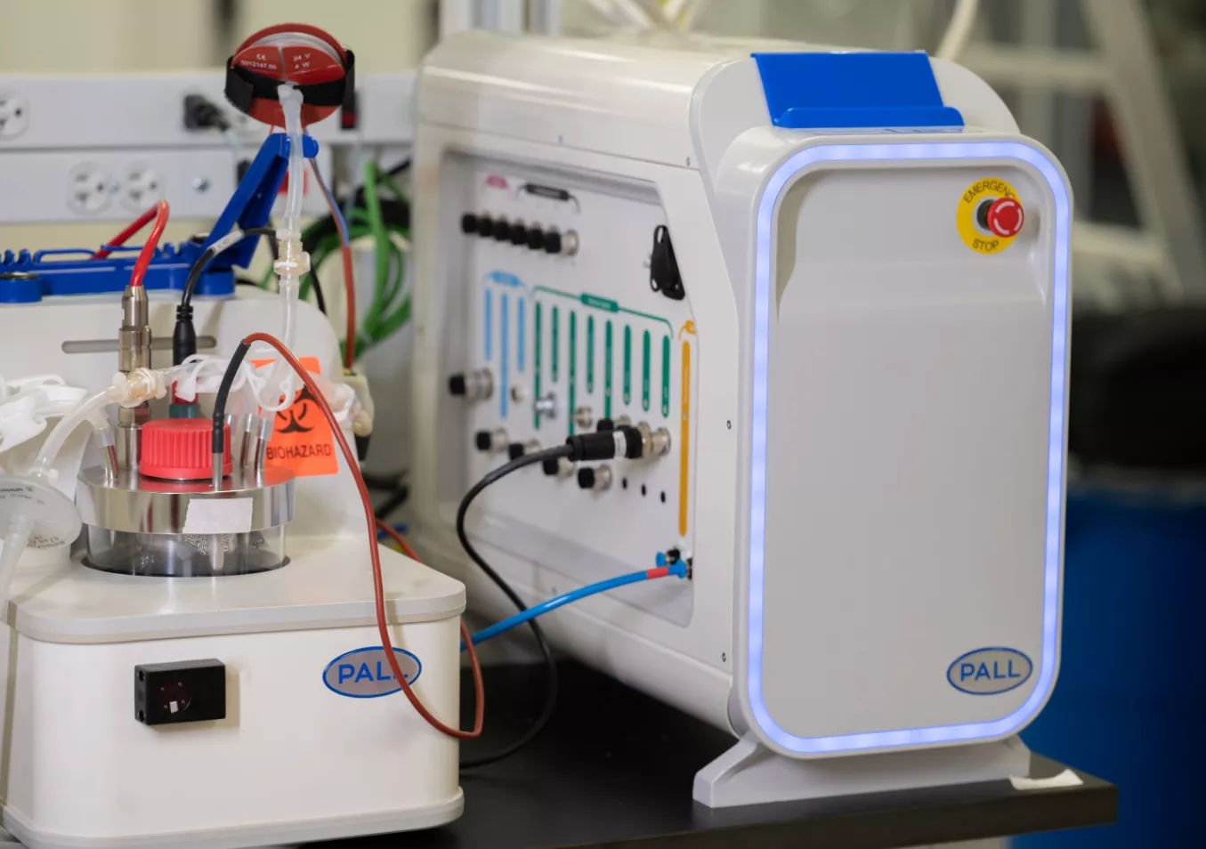
A close-up shot of a modern bioreactor system and control unit from Pall Corporation in a clean laboratory environment.

Defining the In Vivo Fate of EV-Based Therapeutics

Extracellular vesicles represent a compelling class of biologic delivery systems, capable of transporting complex payloads such as nucleic acids and proteins. A definitive understanding of their absorption, distribution, metabolism, and excretion (ADME) profile is a prerequisite for advancing a therapeutic candidate. This characterization directly informs both the safety and efficacy profile by confirming on-target delivery and quantifying potential accumulation in non-target tissues.

Our approach is designed to provide the high-fidelity intelligence for tracking therapeutic delivery that is required for confident decision-making. We design studies that precisely map where your EV therapeutic travels in vivo, how long it persists, and how effectively it delivers its cargo to the intended cellular destination.

Quantitative Bioanalytical Strategies

Accurately measuring EV distribution requires a suite of validated, fit-for-purpose bioanalytical assays capable of overcoming challenges like low signal-to-noise ratios. Franklin Biolabs deploys multiple platforms to build a comprehensive dataset.

  • Nucleic Acid Cargo Quantification: For EVs carrying mRNA, siRNA, or miRNA payloads, validated RT-qPCR assays provide sensitive and specific measurement of cargo delivery at the tissue level.

  • Protein Cargo Quantification: Ligand binding assays are employed to quantify protein payloads, confirming that the vesicle and its cargo arrive at the target site intact.

  • Whole Vesicle Tracking: To understand vector kinetics and localization in real-time, EVs can be labeled with fluorescent or bioluminescent reporters for analysis with in vivo imaging systems.

  • Histology: Tissue-level analysis via Histology provides cellular context for biodistribution data, confirming localization within specific cell types or anatomical structures.

A stylized 3D rendering of a DNA double helix, composed of light-colored spheres on a translucent blue backbone, set against a soft-focus, light blue background.

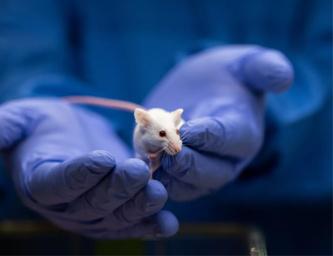
A close-up of a scientist in blue gloves gently holding a small, white laboratory mouse, likely in a research setting.

Lessons from Synthetic Vector Optimization

The development path for EV therapeutics benefits from insights gained in adjacent fields, such as synthetic nanoparticle delivery. For instance, recent work on siloxane-incorporated lipid nanoparticles (PMID: 39354147) demonstrates how targeted chemical modifications can significantly enhance cellular uptake and improve endosomal escape, leading to more potent gene expression in specific tissues.

This principle of rational design to control biodistribution is directly applicable to EVs. Whether through engineering surface proteins or modifying lipid composition, the goal is to optimize the vector for its specific therapeutic purpose. The bioanalytical strategies used to confirm the success of these synthetic vectors provide a robust framework for evaluating next-generation EV candidates.

Integrated Study Design for Regulatory Success

Biodistribution data forms a core component of any successful Investigational New Drug (IND) application. Our studies are designed from the outset with regulatory expectations in mind, conducted in our >100,000 sq ft GxP-compliant facility. This rigorous approach has supported our clients in achieving a 100% IND submission success rate since 2019 (the Franklin Biolabs brand launched in 2024).

Through strategic partnerships, we ensure access to specialized technologies and expertise, creating a cohesive program from vector characterization to final reporting. All in vivo studies adhere to the highest standards of animal welfare, governed by our Animal Welfare Committee and our AAALAC accreditation, with full commitment to the 3Rs (Replacement, Reduction, and Refinement).

Scientific Process Diagram

This content is for informational purposes. For guidance specific to your therapeutic program, please contact our team for a consultation.