Quantitative Analysis of AAV Biodistribution in Cardiac and Skeletal Muscle Tissues

PROVEN INTELLIGENCE ACCELERATING NEXT-GENERATION THERAPIES

Quantitative Analysis of AAV Biodistribution in Cardiac and Skeletal Muscle Tissues

CELL & GENE | RNA | BIOLOGICS

  • Precision Vector Dosimetry for Myotropic Gene Therapies.*
What is the optimal method for quantifying AAV vector copies in dense tissues like cardiac muscle?
The preferred methods are quantitative PCR (qPCR) and droplet digital PCR (ddPCR). Both offer high sensitivity for detecting vector DNA. ddPCR provides absolute quantification without a standard curve, which can be advantageous for reducing variability in complex matrices like homogenized cardiac or skeletal muscle tissue. The choice depends on the specific sensitivity and throughput requirements of the program.
How do you differentiate between vector DNA from transduced cells versus residual vector in the vasculature?
Terminal studies incorporate a whole-body saline perfusion step prior to tissue collection. This procedure effectively flushes the vasculature of circulating vector particles, ensuring that the subsequent DNA analysis primarily quantifies vector copies that have successfully extravasated and are present within the tissue parenchyma or have transduced target cells.
Which AAV serotypes show the highest tropism for skeletal and cardiac muscle?
Serotypes such as AAV8 and AAV9 are widely recognized for their strong tropism for cardiac and skeletal muscle following systemic administration. However, ongoing research continues to identify novel serotypes that can offer equivalent or superior transduction efficiency. Serotype selection is a key variable that must be optimized for each therapeutic candidate based on the target tissue and desired expression profile.
What is the regulatory expectation for biodistribution data in an IND submission for a muscle-targeting AAV?
Regulatory bodies require comprehensive biodistribution data to assess the safety profile of a gene therapy candidate. For a myotropic AAV, this includes robust quantification of vector copies in the target cardiac and skeletal muscles to establish dose-response. It also requires extensive analysis of non-target tissue biodistribution, including gonads, liver, spleen, and brain, to evaluate the potential for off-target effects.

Accurate quantification of adeno-associated virus (AAV) vector biodistribution in cardiac and skeletal muscle is fundamental to developing a viable gene therapy. This analysis directly informs the dose-response relationship, confirms target engagement, and builds the safety profile required for regulatory submission. Franklin Biolabs executes GxP-compliant biodistribution studies using qPCR and ddPCR platforms to deliver precise, IND-ready data packages that de-risk the typical 18-24 month development timeline.

Defining On-Target Efficacy and Systemic Exposure

For AAV-based therapies targeting myopathies or cardiomyopathies, demonstrating preferential delivery to and transduction of muscle tissue is the primary objective. The selection of an appropriate AAV serotype is a foundational step in achieving this goal. Studies show that specific serotypes can provide highly efficient gene transfer to skeletal muscle, establishing stable expression with minimal inflammatory response, a key consideration for long-term efficacy (PMID: 15517544).

However, quantifying on-target delivery is only part of the regulatory requirement. Even with localized administration routes, a significant portion of an AAV dose can distribute systemically. For instance, studies quantifying vector biodistribution have revealed that 60-90% of a dose can disseminate systemically, which has major implications for calculating total body burden and potential immunogenicity (PMID: 37624734). This underscores the need for a comprehensive assessment of non-target tissue biodistribution to construct a complete safety profile.

)

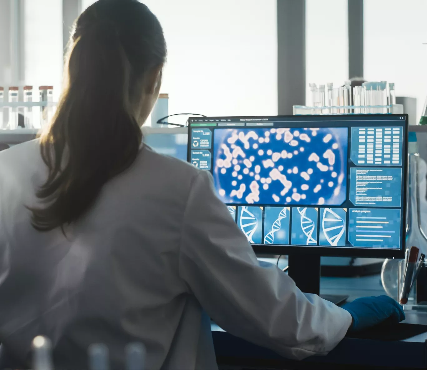
A female scientist in a lab coat analyzes complex biological data, including cellular imagery and DNA models, on a computer screen.

A senior scientist with grey hair is mentoring a diverse group of younger scientists or students in a laboratory setting.

Quantitative Bioanalytical Methodologies

Our >100,000 sq ft facility is equipped to perform sensitive and specific quantification of vector copies from a full range of tissues. The workflow is designed for precision and regulatory compliance.

  • Tissue Processing: Tissues are harvested following rigorous protocols, including saline perfusion to clear the vasculature. Muscle tissues are cryo-pulverized and homogenized to ensure uniform sampling for DNA extraction.

  • Nucleic Acid Extraction: Automated and manual DNA extraction methods are optimized for high yield and purity from fibrous tissues like heart and skeletal muscle.

  • Quantification Platforms: We deploy validated assays on multiple platforms to meet program-specific needs for sensitivity, dynamic range, and throughput.

Platform Key Attribute Optimal Use Case
qPCR High throughput, broad dynamic range Screening large sample sets, established assays
ddPCR Absolute quantification, high precision Low copy number detection, reference standard qualification

“We started collaborating with UPenn Vector core in 2023 and the AAV vector which they manufactured laid a foundation for development of a gene therapy candidate which will enter soon preclinical studies. The key people from UPenn Vector Core joined Franklin Biolabs and our partnership transitioned without interruption from UPenn Vecor Core to Franklin Biolabs Research Vector Division. Franklin Biolabs is our trusted partner in our AAV-vector based gene therapy candidate development and we hope to continue the partnership for years to come.”
— Biotech Partner

IND-Enabling Data for Myotropic AAV Programs

The biodistribution data package is a cornerstone of the CMC and preclinical sections of an IND filing. Our studies are conducted in a GxP environment to ensure data integrity and regulatory acceptance. By providing a clear picture of vector distribution, we help sponsors establish a safe starting dose for first-in-human studies and satisfy agency questions regarding on-target activity and potential non-target tissue toxicities. All study designs adhere to strict animal welfare standards, championing the 3Rs (Replacement, Reduction, and Refinement), and are conducted in AAALAC-accredited facilities to ensure the highest ethical oversight.

Scientific Process Diagram

This content is for informational purposes. For guidance specific to your therapeutic program, please contact our team for a consultation.